Home
/ Long Bone Labeled Endosteum - Long Bone Anatomy Images Stock Photos Vectors Shutterstock - Cartilage synthesis provides for growth in length;
Long Bone Labeled Endosteum - Long Bone Anatomy Images Stock Photos Vectors Shutterstock - Cartilage synthesis provides for growth in length;
Long Bone Labeled Endosteum - Long Bone Anatomy Images Stock Photos Vectors Shutterstock - Cartilage synthesis provides for growth in length;. 31 label the long bone labels design ideas 2020 from www.anatomylibrary99.com structure of long bone although there are many different types of bones in the skeleton, we will discuss the different parts of a specific type of bone in labels may be used more than once. The diaphysis and the epiphysis.the diaphysis is the tubular shaft that runs between the proximal and distal ends of the bone. Match the key terms with the descriptions. When osteoclasts start removing less bone, or osteoblasts start adding more bone, the. Related posts of long bone label chart.
Anatomy/ parts of a long bone vocabulary. The endosteum (plural endostea) is a thin vascular membrane of connective tissue that lines the inner surface of the bony tissue that forms the medullary cavity of long bones. Endosteum occurs beneath the periosteum. On this page, you will find two images i created that illustrate the parts of a long bone and long bone structure. Note that growth plates do not have to be a linear straight line like in the.
Notes Ch 7 Skeleton from www.biologycorner.com Out of the types of bones, the long bones are the most common bones found, and it is. It plays an important role in the healing of fractures by creating new cells necessary for the bone to fuse. Long bone labeled endosteum / labeling portions of a long bone. Cartilage synthesis provides for growth in length; Figure 6.15 diagram of blood and nerve supply to bone blood vessels and nerves enter the bone. Coloring worksheet for this image. They are one of five types of bones: The shaft of a long bone is the diaphysis.
Label the features in your drawings.
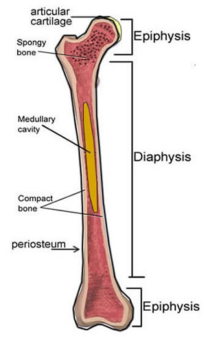
The cavity of long bones consists of red and yellow bone marrow lined with spongy tissue and cancellous bones. The shaft of a long bone is the diaphysis. It covers the loose structures found inside the bone. Endosteum consists of only a cellular layer, which contains bone lining cells, osteoblasts, and osteoprogenitor cells. It is a thin covering that surrounds the medullary cavity. In these labeled examples, a human femur is represented without identifying many of the unique characteristics that help differentiate the femur bone from other bones in the human body. They are composed mostly of compact bone, and are roughly cylindrical in shape with enlarged ends filled with spongy bone. Review of long bone anatomy: Related posts of long bone label chart. Related posts of long bone label chart. Label the features in your drawings. Red marrow end portion of a long bone helps reduce friction at joints site of blood cell formation two membranous sites of 1. The ends of a long bone contain spongy bone and an epiphyseal line.
Label the features in your drawings. The medullary cavity, the hollow spaces in the trabecular (spongy) bone, haversian (osteonic) and volkmann's (perforating) canals in the cortical (compact) bone of the long bones, such as humerus and femur, flat bones, such as ribs 5 and pelvic bones 6, and sesamoid bones, such as patella 10. One of the two rounded ends of the long bone. A long bone has two parts: Where is the endosteum located?
Long Bone Labeled Periosteum Bones Bones Structure Bone Tissue Bone Membranes Gross Anatomy Of A Long Bone Is That It Is Longer Than It Is Wide Examples Are The Tibia from i0.wp.com They are one of five types of bones: This endosteal surface is usually resorbed during long periods of malnutrition, resulting in less cortical thickness. Inside the diaphysis is the medullary cavity, which is filled with yellow bone marrow in an adult. Bones play an important role in anatomy and physiology. It is a thin covering that surrounds the medullary cavity. On this page, you will find two images i created that illustrate the parts of a long bone and long bone structure. Anatomy/ parts of a long bone vocabulary. The diaphysis and the epiphysis.the diaphysis is the tubular shaft that runs between the proximal and distal ends of the bone.
The shaft tends to be cylindrical in form.
The bones shown in the chest and hip region in the labeled human skeleton diagram are the ribs, vertebrae, pelvis, os coxae, sacrum and coccyx. A typical long bone shows the gross anatomical characteristics of bone. The ends that form joints with other bones are the epiphyses (pl.); This endosteal surface is usually resorbed during long periods of malnutrition, resulting in less cortical thickness. The diaphysis is hollow and is made entirely from compact bone. The long bones of the arms are the radius and ulna. Inside the diaphysis is the medullary cavity, which is filled with yellow bone marrow in an adult. Home » anatomy bone labeling » anatomy bone labeling 603 most of the times, we put the labels to show some specific information. The diaphysis is the hollow, tubular shaft that runs between the proximal and distal ends of the bone. Endosteum consists of only a cellular layer, which contains bone lining cells, osteoblasts, and osteoprogenitor cells. Connected to surrounding osteoblasts and osteocytes through. Bones play an important role in anatomy and physiology. The key difference between periosteum and endosteum is that the periosteum consists of an outer fibrous connective tissue layer and an inner osteogenic layer while the endosteum is the thin membranous coating that covers the internal surface of the bone.
Start studying long bone labeled. Review of long bone anatomy: The endosteum is a structure in the middle of bone tissue and bone marrow. Lesson #39 presented long bone anatomy, but let's take a moment to review. Coloring worksheet for this image.
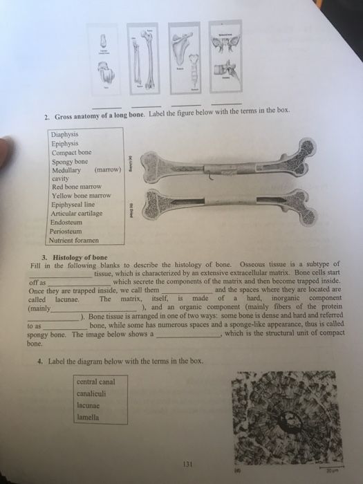
Solved 2 Gross Anatomy Of A Long Bone Label The Figure Chegg Com from media.cheggcdn.com Endosteum lines the inner surface of the medullary cavity of all long bones. This endosteal surface is usually resorbed during long periods of malnutrition, resulting in less cortical thickness. The spongy part of the bone, inner walls of the compact bones and haversian canals. Endosteum occurs beneath the periosteum. Long bones lengthen substantially as a person grows, and have a. Eventually, the cartilage is replaced by bone. Bones play an important role in anatomy and physiology. A epiphysis b diaphysis c articular cartilage d periosteum f compact bone g medullary cavity yellow marrow h endosteum j.
The shaft tends to be cylindrical in form.
Match the key terms with the descriptions. The diaphysis is the hollow, tubular shaft that runs between the proximal and distal ends of the bone. Human skeleton, long bone,epiphysis,diaphysis, spongy bone,compact bone, red bone marrow, yellow bone marrow, medullary cavity, endosteum, periosteum. A epiphysis b diaphysis c articular cartilage d periosteum f compact bone g medullary cavity yellow marrow h endosteum j. Long bone labeled endosteum / labeling portions of a long bone. Long bone labeled illustrations & vectors. Red marrow end portion of a long bone helps reduce friction at joints site of blood cell formation two membranous sites of 1. Eventually, the cartilage is replaced by bone. It is a membrane layer that coats the medullary cavity, bony trabeculae; Label the features in your drawings. A epiphysis b diaphysis c articular cartilage d periosteum f compact bone g medullary cavity yellow marrow h endosteum j. It is a thin covering that surrounds the medullary cavity. The structure of a long bone allows for the best visualization of all of the parts of a bone (figure 1).
The shaft of a long bone is the diaphysis long bone labeled. When osteoclasts start removing less bone, or osteoblasts start adding more bone, the.